Paulingite series | Paulingite-Ca |(Ca0.5,K,Na,Ba0.5)10 (H2O)27-34|[Al10Si32O84] Paulingite-K |(K,Ca0.5,Na)10 (H2O)27-34|[Al10Si32O84] |
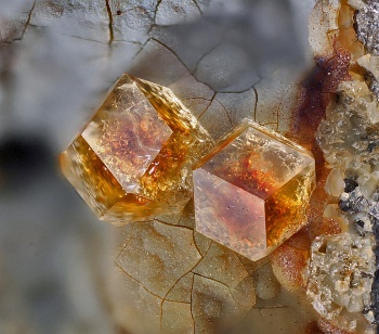
||
Morphology: | ![]() |
||
Isometric. Dodecahedra, {110}, 1 to 5 mm | |||
Physical properties: | |||
Cleavage: none. Hardness: 4 - 4.5. D = 2.085 gm/cm3 (paulingite-Ca) 2.098 gm/cm3 (calc.) ( paulingite-K). Luster: vitreous. Streak: white. |
|||
Paulingite, dodecahedra, Vinarická Hore, near Kladno, Czech Republic. Width of image, 15 mm. (© Volker Betz) | |||
Optical properties: | |||
Color: Colorless to yellowish; colorless in thin section. Isotropic. n 1.482 - 1.484 (paulingite-Ca) n 1.473 - 1.484 (paulingite-K) |
|||
Crystallography: | |||
Unit Cell: | paulingite-Ca | a 35.088 Å (Tschernich and Wise 1982) | |
paulingite-K | a 35.093 Å (Gordon et al. 1966) | ||
Space Group: Im3m | |||
Z = 16 | |||
Names: | |||
Paulingite was described and named by Kamb and Oke (1960). The name honors Professor Linus C. Pauling, Nobel Laureate and Professor of Chemistry at California Institute of Technology. The term paulingite has now been elevated to a series name, covering two species based on the dominant non-framework cation, paulingite-K and paulingite-Ca. The following were proposed as type examples for these species by Coombs et al. (1997): paulingite-K from the original type locality of paulingite along the channel below Rock Island Dam, Douglas County, Washington, USA (Kamb and Oke 1960) and paulingite-Ca from along Three Mile Creek near Ritter, Oregon, USA (Tschernich and Wise 1982). | |||
Crystal structure: | |||
The structure of paulingite was determined by Gordon et al. (1966), confirming the space group Im3m, proposed by Kamb and Oke (1960). The unit cell (a = 35.1 Å) is the largest of all known zeolites, and the framework filling this cell is a complex, though elegant, arrangement of cages and double 8-rings (see PAU). The framework consists of four regular polyhedra or cages and three irregular polyhedra, accounting for the interstices. An alpha-cage (also named lta in the drawingsof PAU) is located at the cell center and at the cell corners. Connected to each of the six single 8-ring faces of the lta are d8R (double 8-ring) and pau polyhedra (see PAU). The sequence of these cages is lta- d8R-pau- d8R-pau- d8R-lta along one edge of the unit cell. Each unit cell contains 672 tetrahedral sites, and the Si,Al distribution throughout these sites appears to be random (Bieniok et al. 1996, Lengauer et al. 1997). 
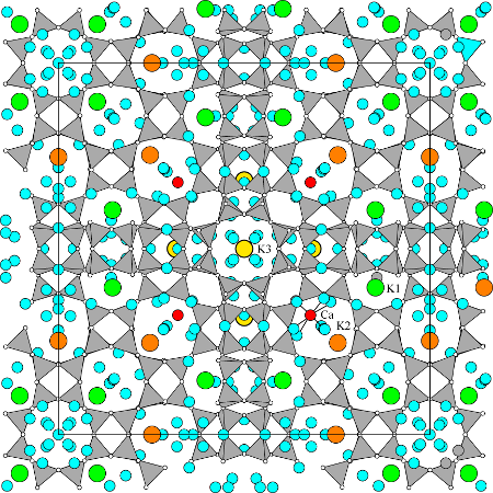
The structure refinement of paulingite-K from Rock Island Dam crystals by Gordon et al. (1966) yielded locations for about 75% of the cations and about 67% of the H2O molecules. More recent refinements by Bieniok et al. (1996) of paulingite-K from Chase Creek, British Columbia and by Bieniok (2000) of paulingite-Ca from Ritter, Oregon, and by Lengauer et al. (1997) of paulingite-Ca from Vinarická Hore, Czech Republic, located more of the 110 to 130 cations and more than 500 H2O molecules per unit cell. Although there is some variation of cation sites from sample to sample, there are also some consistencies. Each of the plg cages contains a site (16 positions per cell) occupied mostly by Ca2+ cations (red in the figure), each surrounded by eight H2O molecules. The K1 site (48 positions per cell, green) is located in each of the irregular 8-ring openings of the pau cage and is coordinated with four framework oxygen anions and two H2O molecules. The K2 sites (24 positions per cell, orange) are outside the plg cages and are generally occupied by K, Ca, and if present, Ba. K2 cations are coordinated with two to six framework oxygen anions, depending on occupancy, and to four H2O molecules. Finally, K3 sites (12 positions per cell, yellow) are within the opr polyhedra connected to grc cages. This site is occupied by K, especially in K-dominant crystals, and is surrounded by six H2O molecules. These four sites account for about 100 cations per cell. The remaining cations, detected by chemical analyses but not located by the refinements, may be randomly distributed among some of the H2O molecule sites not bonded to located cation sites. |
Chemical composition: |
All analyses of paulingite have been made with the electron microprobe, which causes the crystals under the electron beam to fracture and partially collapse. Therefore, placing too much importance on small or even moderate variations in composition may not be warranted. The vacuum and electron beam appears to cause loss of about 60% of the H2O from the excitation volume, giving high partial totals. Commonly the H2O content has been determined by TG or other separate heating methods, and then combined with the microprobe results, and recalculating the full analysis is to 100%. The framework composition is approximately TSi = 0.75, varying only from 0.727 to 0.765. Fe3+ comprises a minor amount, less than 0.07 atoms per formula unit. Non-framework cations are mostly K, Ca, Na, and Ba, with only the first two in dominant amounts. Mg and Sr are detected in most samples but are minor. Three samples have been analyzed for H2O content, which yielded 22.0, 18.50, and 14.30 weight percent (Rock Island Dam, Washington, Gordon et al. 1966; Ritter, Oregon, Tschernich and Wise 1982; and Kladno, Czech Republic, Lengauer et al. 1997, respectively), corresponding to 44.0, 34.3, 27.0 molecules per formula unit. Paulingite easily loses H2O with only moderate heating. Therefore, the variation of H2O content among the three samples may be a result of sample handling and storage, although the differences are still present following crystal structure determinations. For a recent review of the crystal chemistry of the paulingite series see Passaglia et al. (2001). |